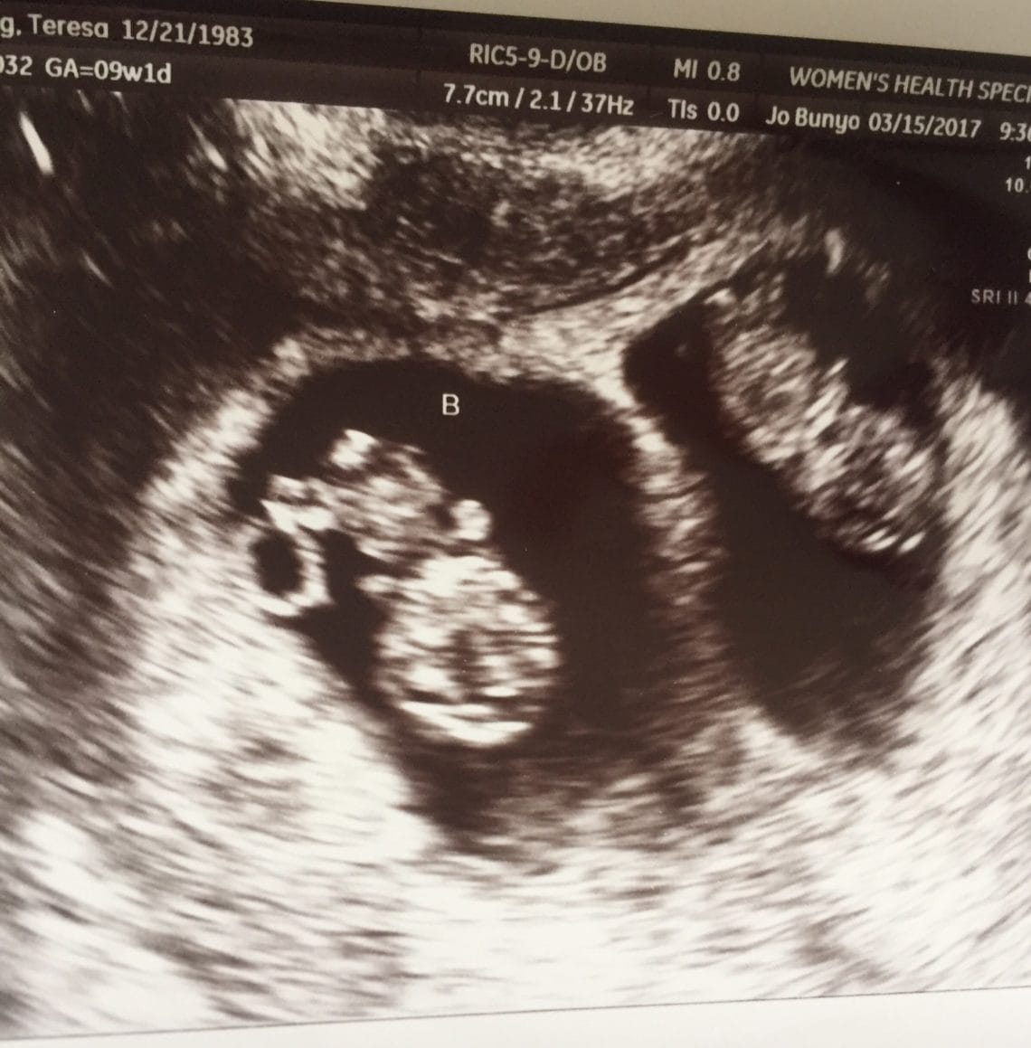
You will need to have a blood test done before coming to Women’s Imaging for your ultrasound scan. What do I need to do to have a risk assessment for my baby? Sound waves are used instead of radiation which makes them safe. Please let reception know at the time of booking.Īn ultrasound scan uses high-frequency sound waves to create images of the inside of your body and baby. You have the option of getting the results of the scan on the day by our consultant. We are able to take some important measurements which allows us to give you an accurate risk assessment for your baby. You will be able to see all of your developing baby If this is extremely painful please let us know.
#12 WEEK SONOGRAM FULL#
Occasionally there is some discomfort from probe pressure on a full bladder or from the vaginal probe manipulation. If you are allergic to latex prior to the vaginal scan or you don’t know then a latex-free cover will be used on the probe. Ultrasound is safe to use throughout your pregnancy.

As well as checking that your baby is growing well and confirming your due date the main aim of the scan includes:

Women’s Imaging conducts a detailed risk assessment for your baby in accordance with the Fetal Medicine Foundation. This scan should be ideally performed between 12 weeks 5 days and 13 weeks 6 days of your pregnancy.


 0 kommentar(er)
